ATP2A1
Protein-coding gene in the species Homo sapiens
| ATP2A1 | |||||||||||||||||||||||||||||||||||||||||||||||||||
|---|---|---|---|---|---|---|---|---|---|---|---|---|---|---|---|---|---|---|---|---|---|---|---|---|---|---|---|---|---|---|---|---|---|---|---|---|---|---|---|---|---|---|---|---|---|---|---|---|---|---|---|
 | |||||||||||||||||||||||||||||||||||||||||||||||||||
| Identifiers | |||||||||||||||||||||||||||||||||||||||||||||||||||
| Aliases | ATP2A1, ATP2A, SERCA1, ATPase sarcoplasmic/endoplasmic reticulum Ca2+ transporting 1 | ||||||||||||||||||||||||||||||||||||||||||||||||||
| External IDs | OMIM: 108730; MGI: 105058; HomoloGene: 7635; GeneCards: ATP2A1; OMA:ATP2A1 - orthologs | ||||||||||||||||||||||||||||||||||||||||||||||||||
| |||||||||||||||||||||||||||||||||||||||||||||||||||
| |||||||||||||||||||||||||||||||||||||||||||||||||||
| |||||||||||||||||||||||||||||||||||||||||||||||||||
| |||||||||||||||||||||||||||||||||||||||||||||||||||
| |||||||||||||||||||||||||||||||||||||||||||||||||||
| Wikidata | |||||||||||||||||||||||||||||||||||||||||||||||||||
| |||||||||||||||||||||||||||||||||||||||||||||||||||


Sarcoplasmic/endoplasmic reticulum calcium ATPase 1 (SERCA1) also known as Calcium pump 1, is an enzyme that in humans is encoded by the ATP2A1 gene.[5][6]
Function
This gene encodes one of the SERCA Ca2+-ATPases, which are intracellular pumps located in the sarcoplasmic or endoplasmic reticula of muscle cells. This enzyme catalyzes the hydrolysis of ATP coupled with the translocation of calcium from the cytosol to the sarcoplasmic reticulum lumen, and is involved in muscular excitation and contraction.[5]
Clinical significance
Mutations in this gene cause some autosomal recessive forms of Brody disease, characterized by increasing impairment of muscular relaxation during exercise. Alternative splicing results in two transcript variants encoding different isoforms.[5] Alternative splicing of ATP2A1 is also implicated in myotonic dystrophy type 1.
ATP2A1 SERCA pumps were very strongly down regulated in amyotrophic lateral sclerosis.[7]
Interactions
ATP2A1 has been shown to interact with:
References
- ^ a b c GRCh38: Ensembl release 89: ENSG00000196296 – Ensembl, May 2017
- ^ a b c GRCm38: Ensembl release 89: ENSMUSG00000030730 – Ensembl, May 2017
- ^ "Human PubMed Reference:". National Center for Biotechnology Information, U.S. National Library of Medicine.
- ^ "Mouse PubMed Reference:". National Center for Biotechnology Information, U.S. National Library of Medicine.
- ^ a b c "Entrez Gene: ATP2A1 ATPase, Ca++ transporting, cardiac muscle, fast twitch 1".
- ^ "UniProt". www.uniprot.org. Retrieved 1 August 2023.
- ^ Mukund, Kavitha; Subramaniam, Shankar (2017). "Co-expression Network Approach Reveals Functional Similarities among Diseases Affecting Human Skeletal Muscle". Frontiers in Physiology. 8: 980. doi:10.3389/fphys.2017.00980. PMC 5717538. PMID 29249983.
- ^ a b Asahi M, Kurzydlowski K, Tada M, MacLennan DH (July 2002). "Sarcolipin inhibits polymerization of phospholamban to induce superinhibition of sarco(endo)plasmic reticulum Ca2+-ATPases (SERCAs)". J. Biol. Chem. 277 (30): 26725–8. doi:10.1074/jbc.C200269200. PMID 12032137.
- ^ Asahi M, Sugita Y, Kurzydlowski K, De Leon S, Tada M, Toyoshima C, MacLennan DH (April 2003). "Sarcolipin regulates sarco(endo)plasmic reticulum Ca2+-ATPase (SERCA) by binding to transmembrane helices alone or in association with phospholamban". Proc. Natl. Acad. Sci. U.S.A. 100 (9): 5040–5. Bibcode:2003PNAS..100.5040A. doi:10.1073/pnas.0330962100. PMC 154294. PMID 12692302.
- ^ Asahi M, Kimura Y, Kurzydlowski K, Tada M, MacLennan DH (November 1999). "Transmembrane helix M6 in sarco(endo)plasmic reticulum Ca(2+)-ATPase forms a functional interaction site with phospholamban. Evidence for physical interactions at other sites". J. Biol. Chem. 274 (46): 32855–62. doi:10.1074/jbc.274.46.32855. PMID 10551848.
- ^ Asahi M, Green NM, Kurzydlowski K, Tada M, MacLennan DH (August 2001). "Phospholamban domain IB forms an interaction site with the loop between transmembrane helices M6 and M7 of sarco(endo)plasmic reticulum Ca2+ ATPases". Proc. Natl. Acad. Sci. U.S.A. 98 (18): 10061–6. Bibcode:2001PNAS...9810061A. doi:10.1073/pnas.181348298. PMC 56915. PMID 11526231.
External links
- Human ATP2A1 genome location and ATP2A1 gene details page in the UCSC Genome Browser.
Further reading
- Baba-Aissa F, Raeymaekers L, Wuytack F, et al. (1998). "Distribution and isoform diversity of the organellar Ca2+ pumps in the brain". Mol. Chem. Neuropathol. 33 (3): 199–208. doi:10.1007/BF02815182. PMID 9642673.
- Callen DF, Baker E, Lane S, et al. (1992). "Regional mapping of the Batten disease locus (CLN3) to human chromosome 16p12". Am. J. Hum. Genet. 49 (6): 1372–7. PMC 1702406. PMID 1746562.
- MacLennan DH, Brandl CJ, Champaneria S, et al. (1988). "Fast-twitch and slow-twitch/cardiac Ca2+ ATPase genes map to human chromosomes 16 and 12". Somat. Cell Mol. Genet. 13 (4): 341–6. doi:10.1007/BF01534928. PMID 2842876. S2CID 1475105.
- Brandl CJ, Green NM, Korczak B, MacLennan DH (1986). "Two Ca2+ ATPase genes: homologies and mechanistic implications of deduced amino acid sequences". Cell. 44 (4): 597–607. doi:10.1016/0092-8674(86)90269-2. PMID 2936465. S2CID 43419264.
- Benders AA, Wevers RA, Veerkamp JH (1996). "Ion transport in human skeletal muscle cells: disturbances in myotonic dystrophy and Brody's disease". Acta Physiol. Scand. 156 (3): 355–67. doi:10.1046/j.1365-201X.1996.202000.x. hdl:2066/24180. PMID 8729696.
- Zhang Y, Fujii J, Phillips MS, et al. (1997). "Characterization of cDNA and genomic DNA encoding SERCA1, the Ca2+-ATPase of human fast-twitch skeletal muscle sarcoplasmic reticulum, and its elimination as a candidate gene for Brody disease". Genomics. 30 (3): 415–24. doi:10.1006/geno.1995.1259. PMID 8825625.
- Odermatt A, Taschner PE, Khanna VK, et al. (1996). "Mutations in the gene-encoding SERCA1, the fast-twitch skeletal muscle sarcoplasmic reticulum Ca2+ ATPase, are associated with Brody disease". Nat. Genet. 14 (2): 191–4. doi:10.1038/ng1096-191. PMID 8841193. S2CID 22726202.
- Bonaldo MF, Lennon G, Soares MB (1997). "Normalization and subtraction: two approaches to facilitate gene discovery". Genome Res. 6 (9): 791–806. doi:10.1101/gr.6.9.791. PMID 8889548.
- Algenstaedt P, Antonetti DA, Yaffe MB, Kahn CR (1997). "Insulin receptor substrate proteins create a link between the tyrosine phosphorylation cascade and the Ca2+-ATPases in muscle and heart". J. Biol. Chem. 272 (38): 23696–702. doi:10.1074/jbc.272.38.23696. PMID 9295312.
- Odermatt A, Taschner PE, Scherer SW, et al. (1998). "Characterization of the gene encoding human sarcolipin (SLN), a proteolipid associated with SERCA1: absence of structural mutations in five patients with Brody disease". Genomics. 45 (3): 541–53. doi:10.1006/geno.1997.4967. hdl:2066/25426. PMID 9367679. S2CID 41989102.
- MacLennan DH, Rice WJ, Odermatt A (1998). "Structure/function analysis of the Ca2+ binding and translocation domain of SERCA1 and the role in Brody disease of the ATP2A1 gene encoding SERCA1". Ann. N. Y. Acad. Sci. 834 (1 Na/K–ATPase a): 175–85. doi:10.1111/j.1749-6632.1997.tb52249.x. PMID 9405806. S2CID 38068023.
- Odermatt A, Becker S, Khanna VK, et al. (1998). "Sarcolipin regulates the activity of SERCA1, the fast-twitch skeletal muscle sarcoplasmic reticulum Ca2+-ATPase". J. Biol. Chem. 273 (20): 12360–9. doi:10.1074/jbc.273.20.12360. PMID 9575189.
- Asahi M, Kimura Y, Kurzydlowski K, et al. (2000). "Transmembrane helix M6 in sarco(endo)plasmic reticulum Ca2+-ATPase forms a functional interaction site with phospholamban. Evidence for physical interactions at other sites". J. Biol. Chem. 274 (46): 32855–62. doi:10.1074/jbc.274.46.32855. PMID 10551848.
- Odermatt A, Barton K, Khanna VK, et al. (2000). "The mutation of Pro789 to Leu reduces the activity of the fast-twitch skeletal muscle sarco(endo)plasmic reticulum Ca2+ ATPase (SERCA1) and is associated with Brody disease". Hum. Genet. 106 (5): 482–91. doi:10.1007/s004390000297. PMID 10914677. S2CID 31756080.
- Daiho T, Yamasaki K, Saino T, et al. (2001). "Mutations of either or both Cys876 and Cys888 residues of sarcoplasmic reticulum Ca2+ATPase result in a complete loss of Ca2+ transport activity without a loss of Ca2+-dependent ATPase activity. Role of the CYS876-CYS888 disulfide bond". J. Biol. Chem. 276 (35): 32771–8. doi:10.1074/jbc.M101229200. PMID 11438520.
- Asahi M, Green NM, Kurzydlowski K, et al. (2001). "Phospholamban domain IB forms an interaction site with the loop between transmembrane helices M6 and M7 of sarco(endo)plasmic reticulum Ca2+ ATPases". Proc. Natl. Acad. Sci. U.S.A. 98 (18): 10061–6. Bibcode:2001PNAS...9810061A. doi:10.1073/pnas.181348298. PMC 56915. PMID 11526231.
- Strausberg RL, Feingold EA, Grouse LH, et al. (2003). "Generation and initial analysis of more than 15,000 full-length human and mouse cDNA sequences". Proc. Natl. Acad. Sci. U.S.A. 99 (26): 16899–903. Bibcode:2002PNAS...9916899M. doi:10.1073/pnas.242603899. PMC 139241. PMID 12477932.
- Pieske B, Maier LS, Schmidt-Schweda S (2002). "Sarcoplasmic reticulum Ca2+ load in human heart failure". Basic Res. Cardiol. 97 Suppl 1 (7): I63–71. doi:10.1007/s003950200032. PMID 12479237. S2CID 22854208.
- Toyoshima C, Asahi M, Sugita Y, et al. (2003). "Modeling of the inhibitory interaction of phospholamban with the Ca2+ ATPase". Proc. Natl. Acad. Sci. U.S.A. 100 (2): 467–72. Bibcode:2003PNAS..100..467T. doi:10.1073/pnas.0237326100. PMC 141018. PMID 12525698.
- v
- t
- e
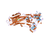
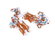
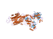
PDB gallery
-
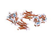 1iwo: Crystal structure of the SR Ca2+-ATPase in the absence of Ca2+
 -
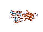
 1kju: Ca2+-ATPase in the E2 State
 -
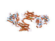
 1su4: Crystal structure of calcium ATPase with two bound calcium ions
 -
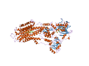
 1t5s: Structure of the (SR)Ca2+-ATPase Ca2-E1-AMPPCP form
 -
 1t5t: Structure of the (SR)Ca2+-ATPase Ca2-E1-ADP:AlF4- form
 -
 1vfp: Crystal structure of the SR CA2+-ATPase with bound AMPPCP
 -
 1wpe:
1wpe: -
 1wpg: Crystal structure of the SR CA2+-ATPase with MGF4
 -
 1xp5: Structure Of The (Sr)Ca2+-ATPase E2-AlF4- Form
 -
 2agv: Crystal structure of the SR CA2+-ATPASE with BHQ and TG
 -
 2by4: SR CA(2+)-ATPASE IN THE HNE2 STATE COMPLEXED WITH THE THAPSIGARGIN DERIVATIVE BOC-12ADT.
 -
 2c88: CRYSTAL STRUCTURE OF (SR) CALCIUM-ATPASE E2(TG):AMPPCP FORM
 -
 2c8k: CRYSTAL STRUCTURE OF (SR) CALCIUM-ATPASE E2(TG) WITH PARTIALLY OCCUPIED AMPPCP SITE
 -
 2c8l: CRYSTAL STRUCTURE OF (SR) CALCIUM-ATPASE E2(TG) FORM
 -
 2c9m: STRUCTURE OF (SR) CALCIUM-ATPASE IN THE CA2E1 STATE SOLVED IN A P1 CRYSTAL FORM.
 -
 2dqs: Crystal structure of the calcium pump with amppcp in the absence of calcium
 -
 2ear: P21 crystal of the SR CA2+-ATPase with bound TG
 -
 2eas: Crystal structure of the SR CA2+-ATPASE with bound CPA
 -
 2eat: Crystal structure of the SR CA2+-ATPASE with bound CPA and TG
 -
 2eau: Crystal structure of the SR CA2+-ATPASE with bound CPA in the presence of curcumin
 -
 2o9j: Crystal structure of calcium atpase with bound magnesium fluoride and cyclopiazonic acid
 -
 2oa0: Crystal structure of Calcium ATPase with bound ADP and cyclopiazonic acid


 | This article on a gene on human chromosome 16 is a stub. You can help Wikipedia by expanding it. |
- v
- t
- e


