Inguinal ligament
Band running from the pubic tubercle to the anterior superior iliac spine
| Inguinal ligament | |
|---|---|
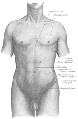 Inguinal ligament is labeled at bottom right. | |
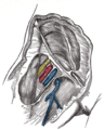
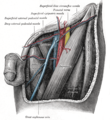
 Structures passing behind the inguinal ligament. Anterolateral view of the right side of the pelvis. | |
| Details | |
| From | Anterior superior iliac spine |
| To | Pubic tubercle |
| Identifiers | |
| Latin | ligamentum inguinale |
| TA98 | A04.5.01.009 |
| TA2 | 2365 |
| FMA | 19855 |
| Anatomical terminology [edit on Wikidata] | |
The inguinal ligament (/ˈɪŋɡwɪnəl/[1][2]), also known as Poupart's ligament or groin ligament, is a band running from the pubic tubercle to the anterior superior iliac spine. It forms the base of the inguinal canal through which an indirect inguinal hernia may develop.
Structure
The inguinal ligament runs from the anterior superior iliac crest of the ilium to the pubic tubercle of the pubic bone. It is formed by the external abdominal oblique aponeurosis and is continuous with the fascia lata of the thigh.
There is some dispute over the attachments.[3]
Structures that pass deep to the inguinal ligament include:
- Psoas major, iliacus, pectineus
- Femoral nerve, artery, and vein
- Lateral cutaneous nerve of thigh
- Lymphatics
Function
The ligament serves to contain soft tissues as they course anteriorly from the trunk to the lower extremity. This structure demarcates the superior border of the femoral triangle.[4] It demarcates the inferior border of the inguinal triangle.
The midpoint of the inguinal ligament, halfway between the anterior superior iliac spine and pubic tubercle, is the landmark for the femoral nerve. The mid-inguinal point, halfway between the anterior superior iliac spine and the pubic symphysis, is the landmark for the femoral artery. The external iliac arteries pass the inguinal ligament posteriorly and inferiorly.
History
It is also referred to as Poupart's ligament, because François Poupart gave it relevance in relation to hernial repair, calling it "the suspender of the abdomen" (French: "le suspenseur de l'abdomen"). It is sometimes termed the Fallopian ligament. Colles' ligament is the reflex ligament and not the inguinal ligament.[5][6]
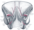
Additional images
-
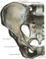 Ligaments of pelvis. Anterior view.
 -
-
 The subcutaneous inguinal ring.
 -
 Femoral sheath laid open to show its three compartments.


-
 The relations of the femoral and abdominal inguinal rings, seen from within the abdomen. Right side.
 -
 The left femoral triangle.
 -
 Posterior view of the anterior abdominal wall in its lower half. The peritoneum is in place, and the various cords are shining through.
 -
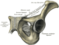
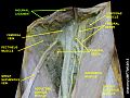 Inguinal ligament
 -
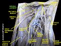 Inguinal ligament


See also
Wikimedia Commons has media related to Inguinal ligament.
- Pelvis
- Apollo's belt: surface features associated with the inguinal ligaments
References
- ^ "Inguinal | Definition of Inguinal by Lexico". Lexico Dictionaries | English. Archived from the original on November 6, 2019.
- ^ "inguinal - WordReference.com Dictionary of English". www.wordreference.com.
- ^ Acland RD (January 2008). "The inguinal ligament and its lateral attachments: correcting an anatomical error". Clin Anat. 21 (1): 55–61. doi:10.1002/ca.20579. PMID 18092366. S2CID 21478857.
- ^ Ryan, Jeffrey M.; Starkey, Chad (2002). Evaluation of orthopedic and athletic injuries. Philadelphia: F.A. Davis Co. ISBN 0-8036-0791-1.
- ^ synd/2633 at Who Named It?
- ^ F. Poupart. Chirurgie complète. Paris, 1695.
External links
- Anatomy figure: 12:03-02 at Human Anatomy Online, SUNY Downstate Medical Center - "Deep muscles of the anterior thigh."
- Anatomy photo:35:os-0107 at the SUNY Downstate Medical Center - "Anterior Abdominal Wall: Osteology and Surface Anatomy "
- Anatomy photo:35:08-0100 at the SUNY Downstate Medical Center - "Anterior Abdominal Wall: The Inguinal Ligament"
- Anatomy image:7179 at the SUNY Downstate Medical Center
- Anatomy image:7431 at the SUNY Downstate Medical Center
- Diagram at gensurg.co.uk
| Authority control databases |
|
|---|


