Inner plexiform layer
Area of the retina
| Inner plexiform layer | |
|---|---|
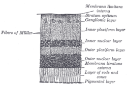 Section of retina. (Inner plexiform layer labeled at right, fourth from the top.) | |
 Plan of retinal neurons. (Inner plexiform layer labeled at left, fifth from the top.) | |
| Details | |
| Identifiers | |
| Latin | stratum plexiforme internum retinae |
| TA98 | A15.2.04.015 |
| FMA | 58704 |
| Anatomical terminology [edit on Wikidata] | |
The inner plexiform layer is an area of the retina that is made up of a dense reticulum of fibrils formed by interlaced dendrites of retinal ganglion cells and cells of the inner nuclear layer. Within this reticulum a few branched spongioblasts are sometimes embedded.[1]
References
- ^ Nolte, John (2002). The Human Brain: An Introduction to Its Functional Anatomy. 5th ed. St. Louis: Mosby. pp. 416–7. ISBN 0-323-01320-1.
External links
- Overview Archived 2010-07-01 at the Wayback Machine at utah.edu
- Histology image: 07902loa – Histology Learning System at Boston University
- v
- t
- e
Anatomy of the globe of the human eye
(outer)
| Sclera |
|
|---|---|
| Cornea |
|

tunic (middle)
| Choroid | |
|---|---|
| Ciliary body | |
| Iris |
| Layers | |
|---|---|
| Cells |
|
| Other |
of the eye
| Anterior segment | |
|---|---|
| Posterior segment |
- Keratocytes
- Ocular immune system
- Optical coherence tomography
- Eye care professional
- Eye disease
- Refractive error
- Accommodation
- Physiological Optics
- Visual perception
| Authority control databases |
|
|---|
 | This article about the eye is a stub. You can help Wikipedia by expanding it. |
- v
- t
- e


