Submucosal plexus
| Submucosal plexus | |
|---|---|
 The plexus of the submucosa from the rabbit. X 50. | |
 | |
| Details | |
| Identifiers | |
| Latin | plexus nervosus submucosus, plexus submucosus, plexus Meissneri |
| MeSH | D013368 |
| TA98 | A14.3.03.042 |
| TA2 | 6728 |
| FMA | 63252 |
| Anatomical terms of neuroanatomy [edit on Wikidata] | |
 |
| This article is one of a series on the |
| Gastrointestinal wall |
|---|
| General structure
|
| Specific
|
| Organs
|
|
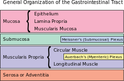
The submucosal plexus (Meissner's plexus, plexus of the submucosa, plexus submucosus) lies in the submucosa of the intestinal wall. The nerves of this plexus are derived from the myenteric plexus which itself is derived from the plexuses of parasympathetic nerves around the superior mesenteric artery. Branches from the myenteric plexus perforate the circular muscle fibers to form the submucosal plexus. Ganglia from the plexus extend into the muscularis mucosae and also extend into the mucous membrane.
They contain Dogiel cells.[1] The nerve bundles of the submucosal plexus are finer than those of the myenteric plexus. Its function is to innervate cells in the epithelial layer and the smooth muscle of the muscularis mucosae.
14% of submucosal plexus neurons are sensory neurons – Dogiel type II, also known as enteric primary afferent neurons or intrinsic primary afferent neurons.[2]
History
Meissners' plexus was described by German professor Georg Meissner.[3]
References
![]() This article incorporates text in the public domain from page 1177 of the 20th edition of Gray's Anatomy (1918)
This article incorporates text in the public domain from page 1177 of the 20th edition of Gray's Anatomy (1918)
- ^ Stach, W (1979). "Die differenzierte Gefäßversorgung der dogielschen Zelltypen und die bevorzugte Vaskularisation der Typ I/2-Zellen in den Ganglien des Plexus myentericus (Auerbach) des Schweins" [Differentiated vascularization of Dogiel's cell types and the preferred vascularization of type I/2 cells within plexus myentericus (Auerbach) ganglia of the pig]. Anatomischer Anzeiger (in German). 145 (5): 464–73. PMID 507375.
- ^ Costa, M (2000). "Anatomy and physiology of the enteric nervous system". Gut. 47 (90004): iv15–9, discussion iv26. doi:10.1136/gut.47.suppl_4.iv15. PMC 1766806. PMID 11076898.
- ^ Haubrich, William S. (February 1999). "Meissner of Meissner's plexus". Gastroenterology. 116 (2): 247. doi:10.1016/S0016-5085(99)70166-6.
External links
- Nosek, Thomas M. "Section 6/6ch1/s6ch1_10". Essentials of Human Physiology. Archived from the original on 2016-03-24.
- Anatomy Atlases – Microscopic Anatomy, plate 10.201
Anatomy of the autonomic nervous system | |||||
|---|---|---|---|---|---|
| Head |
| ||||
| Neck |
| ||||
| Thorax |
| ||||
| Abdomen |
| ||||
| Pelvis |
| ||||
| |||||

Portal:
 Anatomy

| Authority control databases |
|
|---|


